Cataract Surgery

Traditional cataract surgery
Traditional cataract surgery is one of the most frequently performed surgeries and one of the most safe and effective, with predictable outcomes.
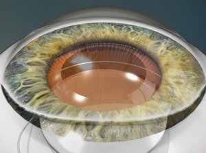
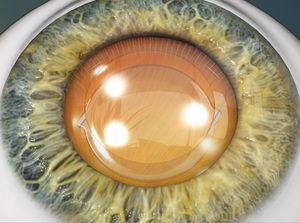
Our surgeons use state-of-the-art technology when performing cataract surgery. To begin, they’ll use a hand-held metal or diamond blade to create an incision in the area where the sclera (the white of the eye) meets the cornea (clear area on top of the eye). Once opened, the surgeon uses special instruments to break up and then gently remove the cataract, which is located right behind the pupil. Next an intraocular lens (IOL) is inserted and implanted, to replace the cloudy natural lens.
Cataract surgery never has to be repeated and the implants last forever. On rare occasions, implants can be exchanged for various reasons, but this is a very rare event.
Take a closer look at your vision post cataract surgery
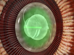
Once the cataract is removed, a replacement lens, called an intraocular lens or IOL, is placed in the eye. The laser can be used to create precise incisions used in lens implantation. Learn more about the cataract replacement lens that may be right for you.


View Video: Cataract Removal (Extracapsular Cataract Extraction Method)

View Video: Cataract Removal (Phacoemulsification Method)

View Video: Intraocular Lenses (IOLs)

