Glaucoma Management
Glaucoma is a group of eye diseases that left untreated can cause optic nerve damage. In glaucoma, eye pressure plays a role in damaging the delicate nerve fibers of the optic nerve. When a significant number of nerve fibers are damaged, blind spots develop in the field of vision. Once nerve damage and visual loss occur, it is permanent. Most people don’t notice these blind areas until much of the optic nerve damage has already occurred. If the entire nerve is destroyed, blindness results.

What causes glaucoma?
Though the exact cause is not fully understood, it is known that it involves the mechanical compression and/or blood flow of the optic nerve. It is commonly associated with elevated eye pressure however, one can develop glaucoma with “normal” eye pressure.
What are the types of glaucoma?
OPEN ANGLE GLAUCOMA
The most common form of glaucoma occurs when the eye’s drainage canals become clogged over time. Also, referred to open-angle glaucoma, the liquid in the eye does not flow efficiently through the eye’s sponge-like drainage system. The fluid builds up and increases the pressure in the eye leading to damage of optical nerve fibers. Most people have no symptoms and no early warning signs.
If open-angle glaucoma is not diagnosed and treated, it can cause a gradual loss of vision. This type of glaucoma develops slowly and sometimes without noticeable sight loss for many years. It usually responds well to medication, especially if caught early and treated.
CLOSED ANGLE GLAUCOMA
More rare but serious type of glaucoma is known as closed-angle or narrow-angle glaucoma. In this type of glaucoma, the iris is not as wide and open as it should be. The outer edge of the iris bunches up over the drainage canals, when the pupil enlarges too much or too quickly. This can happen when entering a dark room. A simple test can be used to see if your angle is normal and wide or abnormal and narrow.
Symptoms of angle-closure glaucoma may include headaches, eye pain, nausea, rainbows around lights at night, and very blurred vision.
How is glaucoma treated?
In most cases glaucoma can be treated with special medicines, medicated eye drops. These medicines lower eye pressure either by reducing the amount of fluid created in the eye or by enabling fluid to flow from the eye. It is important that you administer the medicate eye drops properly. If you have any questions about how to administer eye drops, just ask your ECVA technician or your doctor.
Depending on your case and specific type of glaucoma, your doctor may recommend surgery. The goal of the surgery is to improve the flow of fluid out of the eye, resulting in lowering the eye pressure.

View Video: Argon Laser Trabeculoplasty (ALT) for Glaucoma
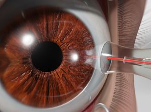
View Video: Laser Cyclophotocoagulation (CPC) for Glaucoma
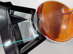
View Video: Selective Laser Trabeculoplasty (SLT)
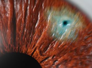
View Video: Laser Peripheral Iridotomy (LPI)
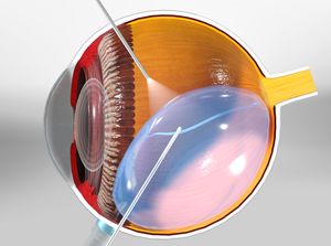
View Video: Vitrectomy
What are the surgical options for treating glaucoma?
LASER TRABECULOPLASTY
A surgery called laser trabeculoplasty is often used to treat open-angle glaucoma. There are two types of trabeculoplasty surgery: argon laser trabeculoplasty (ALT) and selective laser trabeculoplasty (SLT).
Even if laser trabeculoplasty is successful, most patients continue taking glaucoma medications after surgery. For many, this surgery is not a permanent solution. Nearly half of the people who receive this surgery develop increased eye pressure again within five years. Many people who have had a successful laser trabeculoplasty will need more treatment in the future. This treatment may be another laser, more medication or surgery.
SELECTIVE LASER TRABECULOPLASTY – SLT
Selective Laser Trabeculoplasty, or SLT, is a form of laser surgery that is used to lower intraocular pressure in glaucoma.
It is used when eye drop medications are not lowering the eye pressure enough or are causing significant side effects. It can also be used as initial treatment in glaucoma. SLT has been in use for more than 25 years in the United States and around the world.
Who is a candidate for SLT?
Patients who have primary or secondary open-angle glaucoma (the drainage system in the front part of the eye is open) and are in need of lowering of their intraocular pressure (IOP) are eligible for the procedure. Your eye doctor will make the final determination if you are a candidate.
How does it work?
Laser energy is applied to the drainage tissue in the eye. This starts a chemical and biological change in the tissue that results in better drainage of fluid through the drain and out of the eye. This eventually results in lowering of IOP. It may take 1-3 months for the results to appear.
LASER IRIDOTOMY
Laser iridotomy is recommended for treating people with closed-angle glaucoma and those with very narrow drainage angles. A laser creates a small hole about the size of a pinhead through the iris to improve the flow of aqueous fluid to the drainage angle.
TRABECULECTOMY
In trabeculectomy, a small flap is made in the sclera (the outer white coating of your eye). A filtration bleb, or reservoir, is created under the conjunctiva the thin, filmy membrane that covers the white part of your eye. Once created, the bleb looks like a bump or blister on the white part of the eye above the iris, but the upper eyelid usually covers it. The aqueous humor can now drain through the flap made in the sclera and collect in the bleb, where the fluid will be absorbed into blood vessels around the eye.
Eye pressure is effectively controlled in three out of four people who have trabeculectomy. Although regular follow-up visits with your doctor are still necessary, many patients no longer need to use eye drops. If the new drainage channel closes or too much fluid begins to drain from the eye, additional surgery may be needed.
AQUEOUS SHUNT SURGERY
If trabeculectomy cannot be performed, aqueous shunt surgery is usually successful in lowering eye pressure.
An aqueous shunt, or glaucoma drainage device, is a small tube or valve connected to a reservoir (a roundish or oval plate). The plate is placed on the outside of the eye beneath the conjunctiva (the thin membrane that covers the inside of your eyelids and the white part of your eye). The tube is placed into the eye through a tiny incision and allows aqueous humor to flow through the tube to the plate. The fluid is then absorbed into the blood vessels. When healed, the reservoir is not easily seen unless you look downward and lift your eyelid.
A glaucoma valve consists of a small plate with a unique valve system that regulates your eye pressure. Attached to the plate is a tube that drains the fluid out of the eye, thus reducing the eye pressure. The implant is outside the eye, but is covered by the skin of the eye, so it cannot be seen or felt.
There are a variety of implants on the market, but only the Ahmed™ Glaucoma Valve has consistent behavior. The Ahmed™ Glaucoma implant provides effective, long-term control of intraocular press with a high success rate.
