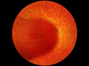
Diabetic Retinopathy
Diabetes mellitus alters the way your body stores and uses dietary sugar. High blood sugar levels can arise from an inadequate amount of insulin (diabetes mellitus type 1) or an insensitivity of the insulin receptors (diabetes mellitus type 2). In either case, these elevated blood sugar levels can cause damage to the small blood vessels throughout your body, and most profoundly to those found in the eye. Damage to blood vessels in the retina is referred to as diabetic retinopathy, the most common type of diabetic eye disease.

View Video: Diabetic Retinopathy
Diabetic retinopathy is a leading cause of blindness in American adults and is preventable. It can arise in anyone with diabetes mellitus (type 1 or type 2) but seems to be related to the length of time one has diabetes and the control of blood sugar levels. It occurs in two forms: non-proliferative and proliferative.
The non-proliferative, or background retinopathy (BDR) is an early stage in which damaged blood vessels can leak blood or fluid into the retina. This fluid can accumulate leading to swelling and deposition of exudate in the retina. If the swelling is central, located at the center of vision-a place called the macula, a symptomatic blurring of vision may occur.
The more severe type, proliferative diabetic retinopathy, begins after prolonged oxygen starvation of the retina. The damaged blood vessels can no longer carry oxygen and nutrients to the retina, so the retina forms new blood vessels. These new vessels are weak and friable (easily broken) and can lead to devastating eye damage. The vessels may break and leak blood throughout the eye, they may form scar tissue in the retina, and they can even result in retinal detachment. Fortunately, early detection can prevent many of the severe complications of diabetic retinopathy.
Symptoms of diabetic eye disease
There are often no symptoms of diabetic retinopathy until significant damage to the retinal blood vessels has taken place. Symptoms at that time include blurred vision, loss of vision, new “spots” or floaters (different from the typical floaters we all get), or shadows. There is no pain or change in the outward appearance of the eye. Because of this lack of symptoms early in the disease, early detection requires an annual visit to your ophthalmologist.
Early detection
It is important to have your eyes examined by an ophthalmologist at the time of diagnosis of diabetes and once a year, every year, after that. The ophthalmologist will dilate your pupils and examine your eyes carefully for any signs of retinopathy. The earliest stages of diabetic retinopathy do not usually need treatment and can respond well to strict blood sugar control. However, if more significant damage is noted, your ophthalmologist may suggest treatment.
Treatment
Historically, treatment of diabetic retinopathy means laser surgery. A strong beam of light is aimed onto the retina to shrink the abnormal vessels or to seal the leaking normal blood vessels. Laser surgery has been shown to reduce the risk of severe vision loss from diabetic retinopathy by over 60 percent but it usually cannot restore vision that has been lost. That is why detection of diabetic retinopathy early is the best way to prevent vision loss.
Modern treatments now involve intravitreal injections of medications such as Avastin and Lucentis, among others. These are classified as anti-VEGF drugs and have been proven to be very safe and effective in treating both macular edema and proliferative diabetic retinopathy. The procedure may need to be performed monthly and in conjunction with laser treatments. It is tolerated very well by patients with appropriate anesthesia.
